Neuritogenesis
Determine the function of membrane-bending proteins in cortical neuron migration and process outgrowth
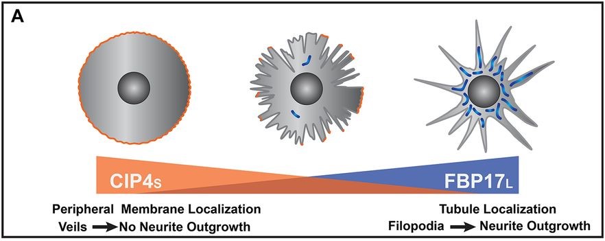
Figure adapted from Taylor et al 2019, depiction of opposing functions of CIP4 and FBP17
To date, our lab has focused on the two F-BAR proteins CIP4 and FBP17. While these proteins are structurally very similar, they have striking and opposing effects on developing neurons. CIP4 localizes to the periphery of developing neurons, leading to broad expanses of lamellipodia. FBP17 localizes to endocytic tubules, and results in neurons with excess neurite formation.
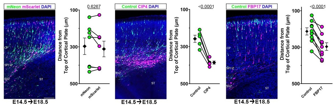
Figure adapted from English et al 2025. Briefly, each image shows migration of two distinct populations. Green neurons are controls, essentially wildtype. Pink neurons are expressing either no manipulation (left image), CIP4 (middle image) or FBP17 (right image). Graphs are demonstrating spread and reliability of data across multiple experiments.
We have investigated the roles of these proteins in developing mouse cortex via the technique in utero electroporation. Sustained expression of either CIP4 or FBP17 leads to an overall breakdown of migration. Both maintain the morphological changes seen in vitro (rounded cells for CIP4, excess neurites for FBP17) , but sustained expression of either is sufficient to halt migration before neurons arrive at the cortical plate.